 |
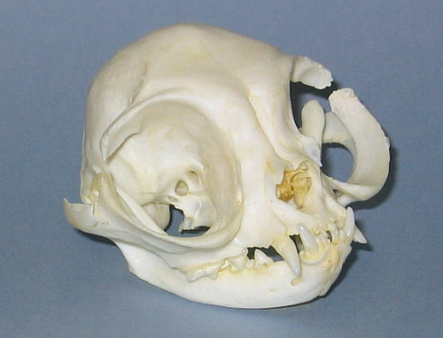
| Brachycephalic (short-headed or round shaped respectively) |
 |
| Mesocephalic (normal shaped or mesatocephalic respectively) |
Possible health problems in extreme typed, flat faced, Persians.
Brachycephaly
(short-headed / A disproportionate shortness of the
head)
Brachycephalic airway syndrome
Excessive
tearing
(Tear duct overflow)
Ulcerated Cornea
(scratch
or rupture of the eyeball)
Stenotic Nares
(Small
nostrils)
Dermatitis
(skin
infection)
Malocclusions
(abnormal
positions of teeth)
Cleft palates
Dystocia
(difficulty
giving birth)
Solution
Brachycephaly (short-headed / A disproportionate shortness of the head)
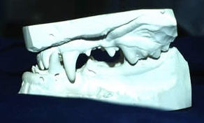
In order to enlighten the dark labyrinth of the nasal cavity, a comparative examination of brachycephalic (short-headed or round shaped respectively) and mesocephalic (normal shaped or mesatocephalic respectively) cats was carried out. The study was based on 26 Persian and 18 domestic cats in total.
Because the animals displayed
different stages of brachycephaly, and in order to adequately describe the
functional-anatomical alterations, the 26 examined Persian cats were divided
into four different categories by subjective criteria. This study presents the
first complete cross-sectional anatomic descriptions of the nasal and paranasal
cavities in brachycephalic cats, and – at least regarding some areas – even
in mesocephalic domestic cats. Because the nasolacrimal drainage system of the
cat was hitherto not described using computed tomography, this gap regarding
functional anatomy and clinical diagnosis was thus closed.
The main problems when looking at the feline nose regarding the complex
alterations in brachycephaly arise from the highly shortened facial bones and
the resulting dislocation of nasal structures caused by the dorso-rotation of
the teeth. Concomitant with increased stages of brachycephaly, the nares and the
nasal entry get narrower; the rostral ending of the respiratory duct, the nasal
conchae and the whole ethmoidal bone are pushed into an increased upright
position; and the nasolacrimal drainage system is characterized by an increased
angle and a steeper course. Nasal conchae material is pushed into the
respiratory duct in some animals of the stage III category and thus hinders
respiratory air flow. Additional paranasal cavities (Sinus conchales of the
ectoturbinals 2 and 3) are developed, while the upper ethmoidal conchae are
pushed up towards the frontal sinuses by the compression processes within the
lower aspects of the nasal cavity.
The literature survey presents the nose as a functional system, and based on
this the present study focuses on all important diagnostic cross-sectional
anatomic problems in brachycephalic and mesocephalic cats. Besides the nasal
conchae, the nasal ducts and the paranasal sinuses, even the mucous membranes,
the vasculature, regional lymph nodes and the nasolacrimal drainage system are
considered. The results presented on the complex of ‘brachycephaly in
cats’ suggest that brachycephalic animals should be classified into four
categories (I – V) regarding objectionable structural criteria and use this
classification interpreting the law of animal welfare (‘pain breed’ in terms
of paragraph 11b). Cats of the category IV seem to fulfil the corpus delicti of
‘pain breed’. Breeders of brachycephalic breeds should refrain from breeding
with animals even of category III. According to the present study, even animals
of category I and II display alterations due to dorso-rotation (slight narrowing
of the nasal entry, low grade angling of the respiratory duct and the
nasolacrimal drainage system, development of additional paranasal sinuses). But
- in the hands of conscientious breeders - the latter add to the variety of
feline breeds and are not objectionable from the veterinary point of view
Source:
http://www.diss.fu-berlin.de/2007/442/indexe.html
|
|
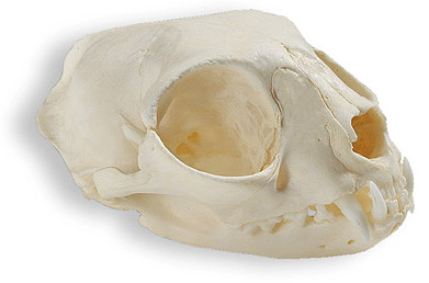
| Brachycephalic (short-headed or round shaped respectively) |
|
|
| Mesocephalic (normal shaped or mesatocephalic respectively) |
Brachycephalic airway
syndrome
Brachycephalic airway syndrome is defined as partial blockage of airflow through
the upper respiratory tract, which consists of the area from the nostrils to the
throat. It is seen more frequently in animals with typical brachycephalic
('pug-nosed') conformation consisting of a flat face, round head, and thick,
short neck.
Brachycephalic airway syndrome typically occurs in specific breeds of dogs (such
as the bulldog, pug, and boxer) and cats (such as the Persian and Himalayan).
Brachycephalic airway syndrome is caused by compressed or narrowed air passages,
which are typical of dogs or cats with brachycephalic conformation. In addition,
diseases of the respiratory system, environmental factors (such as stress,
exercise, or extreme temperatures), and some types of systemic disease can
intensify the signs associated with brachycephalic airway syndrome.
The signs of brachycephalic airway syndrome include noisy or open-mouth
breathing, snoring, panting, exercise intolerance, vomiting, and difficulty
eating. An exaggerated movement of the animal's abdomen during respiration is
seen commonly in more severely affected animals.
The tissues lining the upper respiratory tract also may be damaged or inflamed
because of the large amount of force that is generated when the animal breathes.
The muscles involved in respiration can be abnormally large because of the
increased effort required for the process of breathing.
Brachycephalic airway syndrome is diagnosed by medical history and physical
examination in a brachycephalic breed. It is diagnosed by observing the
characteristic brachycephalic conformation and detecting a partial obstruction
or blockage in airflow along the upper respiratory tract. A laryngoscope (an
instrument used to look into the throat and windpipe) may be used to identify an
obstruction that is not detected easily on physical examination. Mild sedation
of the animal to facilitate examination of the throat area may be needed to
identify problems in the soft palate or throat (pharynx). Radiographs (X-rays)
may be necessary.
The goal in the treatment of
brachycephalic airway syndrome is to make it easier for the animal to breathe.
Some treatment options include exercise restriction (especially when it is hot),
obesity prevention, and elimination of stress. Surgery may be required for
animals suffering from severe respiratory distress.
Most animals are able to compensate with appropriate treatment and management.
However, the long-term prognosis can become poor over time because the signs of
this disease worsen as the animal ages.
Source:
http://www.petalia.com.au/templates/storytemplate_process.cfm?specie=Dogs&story_no=1875
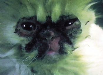
Excessive tearing (Tear duct overflow / Epiphora)
Epiphora is an abnormal overflow of tears down the face that results from
either obstruction of tear drainage through the nasolacrimal (tear duct) system
or overproduction of tears that overwhelms the normal drainage system. The
overproduction of tears is most often a reflex, activated to expel irritating
material from the surface of the eye or when significant irritation develops
inside the eye. Although uncommon, epiphora may also result from overactive
lacrimal (tear) glands and be unassociated with any source of irritation.
Tears are continuously produced on the eye in most animals. With each blink of
the eyelids, tears are pushed along the outer aspect of the eyelids towards the
nose.
While tears are typically
colorless, they can dry to a dark red-brown-black crust, especially in the cat.
Chronic tearing can also result in a brown to rust-colored staining of the hair
around the eyes and face of animals. This is believed to be due to porphyrins or
other pigment-like substances present in the tears.
Tears that spill over onto the face can also be irritating to the skin of the
face. Moisture and bacterial build-up in that area aggravates this irritation.
Epiphora can be caused by numerous conditions.Inefficient drainage of tears from
partial closure of the drainage openings, increased kinking of the drainage duct
in the nose, or wicking of tears onto hairs present in the crease where the
eyelids meet. This condition is most common in the flat faced, long-haired
breeds of cats, particularly the Persian and Himalayan breeds.
As a result of their short and concave underlying facial bone structure, the
lacrimal sac and the nasolacrimal duct of most Persian cats is blocked at the
lacrimal puncta, causing an excessive coagulation of debris and an overflow of
tears from the lacrimal glands: Excessive tearing is a common characteristic of
Persian cats and is caused by abnormal drainage of tears and may result in
epiphora (Faculty of Cornell Feline Health Center 1997). The two lacrimal puncta
are the small openings to the canaliculi (ducts) leading to the lacrimal sac.
The nasolacrimal duct drains the sac into the nose. The ducts of the lacrimal
system are already very small in felines and the facial conformation of
extremely short-nosed, large-eyed cats, namely Persians, is the single most
common cause of occlusion (blockage) of the lacrimal system and resulting
abnormal drainage of tears. Consequently, because of the epiphora and the
blocked lacrimal system, Persian cats suffer from chronic eye infections.
Bacteria build up in the obstructed lacrimal passages and thrive on the debris
deposited from the coagulated tears, inducing chronic conjunctivitis, with
characteristic symptoms such as a brown, mucus-like discharge from the eye,
blinking, and an exposed third eyelid. In severe cases, the conjunctiva is
swollen red (Faculty of Cornell Feline Health Center 1997). It is important to
note, however, that conjunctivitis is not painful (Carlson and Giffin 1995).
Sources:
http://www.petplace.com/cats/epiphora-in-cats/page1.aspx
http://www.simbakui.com/thesis.htm
Ulcerated Cornea
(scratch or rupture of the eyeball)
An ulcerated cornea is a scratch or rupture of the eyeball. Because extreme
typed Persians have such large eyes, it is very easy for adults and kittens to
poke or scratch their eyeball, or become scratched in the eyeball by another cat
or animal in the house. When a cat becomes afflicted with an ulcerated cornea,
infection usually follows and the cat can become blind. Treatment is
possible through optical antibiotics but if the scratch is deep enough, a
veterinarian will have to surgically close the eyelids for a period of 2-6 weeks
in order to allow the eye to heal. The scratch will heal if properly cared for,
but a scar will always be present causing possible obstruction of vision and the
inability for the cat to be shown. In some cases, such as ones involving deep
scratches and untreated ulcers, the eyeball will have to be permanently removed
from the Persian's face. If not properly cared for, or left un-treated, the
ulcer will cause the cornea to pull away from the retina, causing the eyeball to
show a black "tumor-like" abscesses on the surface which cannot be
corrected even with proper veterinary care. Signs of ulcerated cornea include
digging at the eye, pussy/hardly opened eyes, a blackened eyeball, and frequent
infection. If any of these symptoms occur, please contact a veterinarian
immediately for advice and treatment.
Stenotic Nares (Small
nostrils)
Small nostrils, pinched nostrils, slit nose holes... are more correctly called
Stenotic Nares. Stenotic nares are most often seen in the brachycephalic breeds
of cats and dogs... Persians, Himalayans... Boxers, Pugs, Lhasa Apsos... the
breeds with pushed in faces.
Any Persian cat breeder can produce a cat with this problem.
Small nostrils are genetic, however the process of inheritance is
polygenic, indicating multiple genes are involved. What does this mean? It means cats with wide open nostrils can produce kittens with small nostrils. And cats with small nose holes can produce kitties with normal nose openings. So regardless of how conscientious a breeder may be about never using breeding stock with small nostrils, the day may come when any breeder may need to deal with the problem of a cat with stenotic
nares.
Usually, the wing of the nostril blocks the opening and when the cat breathes
in, the wing is pulled in further and blocks the opening even more.
This can cause the
cat to struggle to breath properly. Severely affected cats can be heard
breathing across the room. The negative pressures caused by the added effort of
breathing can also cause deformations of the soft palate and trachea formation.
Affected cats often eat poorly and show stress when playing or being very active.
An operation that enlarges the nose holes can result in remarkable improvements
and a more comfortable life for the cat. While enlarging the holes can be
accomplished with conventional scalpel techniques, the use of laser surgery
often obtains superior results.
Source:
http://www.showcatsonline.com/x/enlarging_small_nostrils_on_a_persian_cat.htm
Dermatitis (skin
infection)
 Persians
are prone to watery, drippy eyes. The flatter and more
extreme the nose, the more tearing will be produced and very few extreme typed
Persians do not have drippy eyes. Not only does this leave your home stained
from the brown eye snot, it can be unhealthy for the cat if not cleaned daily.
With the fold above the nose hiding a deep cavity, daily maintenance is required
to keep the eye snot from dripping all over your home and also to keep the cat's
face clean and healthy. Unfortunately, the cavity above the nose (it's called a
nose break) fills up with the drippy tears daily and with the lack of air
circulation to this area of the face, it is very possible for the flat faced
Persian to get pyoderma (skin infection) or fungus growing in the fold and
cavity.
Persians
are prone to watery, drippy eyes. The flatter and more
extreme the nose, the more tearing will be produced and very few extreme typed
Persians do not have drippy eyes. Not only does this leave your home stained
from the brown eye snot, it can be unhealthy for the cat if not cleaned daily.
With the fold above the nose hiding a deep cavity, daily maintenance is required
to keep the eye snot from dripping all over your home and also to keep the cat's
face clean and healthy. Unfortunately, the cavity above the nose (it's called a
nose break) fills up with the drippy tears daily and with the lack of air
circulation to this area of the face, it is very possible for the flat faced
Persian to get pyoderma (skin infection) or fungus growing in the fold and
cavity.
Skin
diseases often
cause itching, inflammation and painful open sores or ulcers and lead to the
infection of broken skin and sores by bacteria. Skin disease can be a major
welfare problem for the animals and stressful for the owner. Veterinary
textbooks make it clear that pedigree dog & cat breeds are predisposed to a
number of distressing skin complaints. P B Hill, of the Royal (Dick) School of
Veterinary Studies, University of Edinburgh, a specialist in veterinary
dermatology, states that skin diseases ‘show striking breed predilections.’30
In Hill’s textbook, Small animal dermatology. A practical guide to the
diagnosis and management of skin diseases in dogs and cats (2002), breed related
skin diseases are listed for 56 dog breeds plus the Persian cat
Dogs and cats bred to have loose or drooping skin are most likely to suffer from
skin fold dermatitis (intertrigo). Skin fold dermatitis is caused by friction
between skin surfaces, and leads to ulceration, exudation of pus and fluid,
surface infection and foul odour. Sores due to intertrigo are found in the areas
where there are skin folds in the different affected breeds. The Boston Terrier,
Bulldog, Pekinese, Pug and Persian cats are affected in the skin folds on their
faces.
Fold dermatitis with associated pyoderma is one of the conditions that results
from selection by breeders and a demand from the public for particular features,
in this case skin wrinkling, heavy facial folds, or a "corkscrew"
tail. These disorders are directly related to the conformation or standards for
the breed. Although these conditions have in many cases become so common that
they are accepted as normal for the breed, they can still cause serious physical
problems and discomfort for the animal.
A severe form of the dermatitis is reported in Persians - it is known as
idiopathic Persian facial dermatitis - some veterinary surgeons call it 'dirty
face'. It can be very difficult to manage. The skin of such cats shows a black
waxy material on the hairs in a symmetrical pattern on the face, but
particularly the chin and around the eyes.
Sources:
http://www.advocatesforanimals.org.uk/pdf/Thepriceofapedigree.pdf
http://www.upei.ca/~cidd/Diseases/dermatology/fold%20dermatitis.htm
http://www.rarebreedcattery.com/health.html
Malocclusion
abnormal
positions of teeth
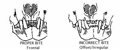 Malocclusion
refers to an abnormal tooth alignment. The short, wide heads cause increased tooth crowding, causing the teeth to rotate or overlap
causes a higher incidence of periodontal disease. Abnormalities of occlusion in
cats include: prognathic malocclusions, brachygnathic malocclusions,
wry mouth rostromesial inclination of the maxillary canine tooth, and
lingual displacement of mandibular canine teeth. Prognathic malocclusions occur
most frequently in Persians. Occasionally, excessive dolichocephalism in Siamese
cats may result in brachygnathic malocclusions. Wry mouth is a skeletal
malocclusion in which one or more of the four quadrants of the jaw grow
disproportionately to the others, resulting in improper alignment of the
midlines of the dental arcades. Wry mouth occurs most frequently in extreme
flat-nosed brachycephalic cats that have a malpositioned mandibular canine tooth
that remains exposed when the mouth is closed. his is the worst of all bite
problems. Wry mouth can cause a severe handicap when it comes tograsping items
or chewing food, and gives a very lopsided appearrance.
Malocclusion
refers to an abnormal tooth alignment. The short, wide heads cause increased tooth crowding, causing the teeth to rotate or overlap
causes a higher incidence of periodontal disease. Abnormalities of occlusion in
cats include: prognathic malocclusions, brachygnathic malocclusions,
wry mouth rostromesial inclination of the maxillary canine tooth, and
lingual displacement of mandibular canine teeth. Prognathic malocclusions occur
most frequently in Persians. Occasionally, excessive dolichocephalism in Siamese
cats may result in brachygnathic malocclusions. Wry mouth is a skeletal
malocclusion in which one or more of the four quadrants of the jaw grow
disproportionately to the others, resulting in improper alignment of the
midlines of the dental arcades. Wry mouth occurs most frequently in extreme
flat-nosed brachycephalic cats that have a malpositioned mandibular canine tooth
that remains exposed when the mouth is closed. his is the worst of all bite
problems. Wry mouth can cause a severe handicap when it comes tograsping items
or chewing food, and gives a very lopsided appearrance.
A maxillary canine tooth that is displaced rostrally may occlude traumatically
with the mandibular canine, causing labial displacement of the mandibular canine
tooth and secondary traumatic occlusion of the mandibular canine with the lip.
This is known as rostromesial inclination of the maxillary canine tooth. This
traumatic occlusion by the mandibular canine tooth may cause a lesion resembling
an eosinophilic granuloma. Retained deciduous mandibular canine teeth may result
in lingual displacement of the permanent mandibular canine teeth resulting in
traumatic occlusion with the hard palate. When abnormalities of occlusion result
in traumatic occlusion including soft tissue trauma and tooth-to-tooth trauma
treatment in the form of exodontia, orthodontia or endodontia is recommended.
If the molar teeth are maloccluded, they can develop “points,” sharp edges
that result from the uneven wear of the teeth. These points can cut into the
inner cheeks and tongue. This is not only very painful, but also can lead to
infection in these areas.
Prognathia (undershot):
 Prognathia (undershot) is found when the mandible is longer relative to the
maxilla. It is identified on oral examination by finding the mandibular incisors
in contact with or rostra to the maxillary incisors. In brachycephalic dogs and
Persian cats, it is considered a normal breed characteristic. Despite being seen
to varying degrees, it rarely requires any specific treatment.
Prognathia (undershot) is found when the mandible is longer relative to the
maxilla. It is identified on oral examination by finding the mandibular incisors
in contact with or rostra to the maxillary incisors. In brachycephalic dogs and
Persian cats, it is considered a normal breed characteristic. Despite being seen
to varying degrees, it rarely requires any specific treatment.
Sources:
http://www.merckvetmanual.com/mvm/index.jsp?cfile=htm/bc/20202.htm
http://www.vin.com/VINDBPub/SearchPB/Proceedings/PR05000/PR00355.htm
http://www.cfa.org/articles/health/dental.html
http://www.veterinarypartner.com/Content.plx?P=A&A=164&S=2
http://agrowebcee.net/subnetwork/faw/reports2/CoE.pdf
http://ms.yccd.edu/vet01/vett53a/vett53Anotes/notes/notes24.htm
Cleft palates
A cleft palate is an opening between the mouth (oral cavity) and the nose (nasal
cavity) that occurs when the tissues separating these two cavities do not grow
together properly. This birth defect can occur in the lip (“primary
cleft palate”, “cleft lip”, or “harelip”) or along the roof of the
mouth (“secondary cleft palate”). Within the mouth, the cleft can
extend along the bony portion (hard palate), the flexible portion that is used
in swallowing (soft palate), or both.
Brachycephalic breeds such as Persians are reported to have about 30% risk.
Signs:
There is a visible split in the roof of the mouth or the soft palate. This results in food material (particularly fluids) passing into the nasal cavity and the following signs :
Bronchopneumonia accounts for 30% of deaths due to cleft palate.
Brachycephalic breeds are
short-faced because of abnormal pituitary/hypophysis glands. This "master
gland" affects all other endocrine glands and all hormones, directly or
indirectly. Including those that affect cleft palate. Perhaps the defect that
causes a flat face with pushed-in nose and undershot jaw is side-by-side with
the defect that directly or indirectly interferes with normal midline closure in
the embryo just before birth or earlier in gestation.
Sources:
http://www.provet.co.uk/health/diseases/cleftpalate.htm#c
http://siriusdog.com/cleft-palate-sevin-congenital-dog.htm
http://www.vin.com/proceedings/Proceedings.plx?CID=WSAVA2006&PID=16079&O=Generic
Dystocia (difficulty giving birth)
Gunn-Moore DA,
Thrusfield MV. 1995 Feline Dystocia : prevalence, and association with cranial
conformation and breed.Vet Rec. 136(14):350-3:
The litter prevalence of feline dystocia was investigated using a questionnaire
survey of cat breeders. Information was obtained on 2928 litters, from 735
queens. Dystocia was reported to have occurred in 5.8 per cent of litters. The
level of dystocia in individual breeds ranged from 0.4 per cent of litters born
in a large colony of cats of mixed breeding, to 18.2 per cent of litters in the
Devon rex. Pedigree litters were at significantly higher risk than litters of
cats of mixed breeding (odds ratio: 22.6). Relatively high levels of dystocia
were identified in Siamese-type, Persian and Devon rex litters, whereas cats of
mixed breeding showed a relatively low litter prevalence. Dolicocephalic and
brachycephalic types were found to have significantly higher levels of dystocia
than mesocephalic cats.
Malposture is of most importance in relation to the position of the head.
Brachycephalic cats may have difficulty at the point where the foetal head first
engages the opening of the maternal pelvis. The lack of a wedge-shaped muzzle
increases the risk of the head becoming deflected to one side, downwards between
the forelegs, or onto the chest. Occasionally, one or both forelegs may lie back
along the body, and in posterior or tail-first presentation one or both hind
legs may be retained forwards alongside the body to give the breech posture. All
of these situations may give rise to either a temporary delay and necessitate
extra efforts by the cat or, at worst, result in complete obstruction.
Sources:
http://www.michvet.com/library/surgery_dystocia.asp
http://intl.elsevierhealth.com/e-books/pdf/974.pdf
http://veterinaryrecord.bvapublications.com/cgi/content/abstract/136/14/350
www.dr-addie.com/Conditions.htm
http://www.petdoctoronline.com/articles/feline_part.html
Solution:
Guidelines for the revision of breeding policies:
- set limits to the shortness of skull, respectively nose, so that breathing difficulties and blockage of lachrymal ducts are avoided, as well as disposition to birth difficulties
- prevent the occurence of:
-abnormal positions of teeth ( brachygnathia) to avoid difficulties in feeding and caring for the newborn;
-abnormal size and form of eyes or eyelids to avoid irritation, inflammation and degeneration as well as prolapse of eyes;